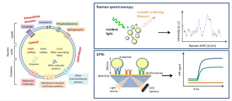
Extracellular vesicles are nanoscaled membrane vesicles released by multiple cell types under both physiological and pathological conditions. Together with microparticles and exosomes, they are considered interesting cargoes for bioactive molecules useful in the intercellular paracrine communication. Their remarkable potentialities in diagnostics and therapeutic treatment of multiple complex diseases such as degenerative diseases of permanent tissues (i.e. central nervous system and muscles) make their deep characterization urgent. LABION is currently working at the biochemical characterization of extracellular vesicles released from cultured mesenchymal stem cells and present in accessible biofluids by Raman spectroscopy and of circulating extracellular vesicles involved in neurodegenerative diseases by SPRi.  – Raman spectroscopy is an interesting new approach for vesicle characterization as it allows the evaluation of their overall chemical composition from a tiny volume of sample avoiding vesicle labeling. We propose the use of Raman spectroscopy both as rapid quality check tool of vesicles to be used for in vitro or in vivo experiments and as biochemical characterization method.
One of Labion ongoing projects is aimed at the characterization of vesicles isolated by ultracentrifugation from the conditioned medium of human mesenchymal stem cells cultured as monolayer in vitro in serum-deprived conditions. We have demonstrated the ability of the method to successfully detect the main constituents of the isolated vesicles (Gualerzi et al, 2017, Sci Rep). More recently, we have also demonstrated that Raman Spectroscopy represents a valuable and quick tool to assess purity of extracellular vesicle preparations and predict their functionality (Gualerzi et al, 2019, JEV).
The Raman analysis was also successfully applied to the vesicles isolated from the serum of patients affected by Parkinson’s disease demonstrating that the biochemical modifications of vesicles are related to the disease progression (Gualerzi et al., 2019, Nanomedicine)
– SPRi (Surface Plasmon Resonance imaging) is an analytical technique able to detect variations in the mass adsorbed on top of a gold–coated chip, occurring in a layer of about 200 nm from the surface, as a result of interaction between probe molecules immobilized on the chip and capture target molecules.
We propose the use of SPRi to facilitate extracellular vesicles applied research, allowing the study of the interactions between vesicles and specific biomolecules. Taking advantage of SPRi technology, we aim to simultaneously detect several markers on extracellular vesicles’ membrane with high sensitivity using the mass of the membrane as a source of amplification of the signal.
In particular, we have designed a SPRi-based platform for the detection and characterization of multiple vesicle populations of neural origin directly in the blood, paving the road toward more precise clinical studies on the use of vesicles as potential biomarkers of neurodegenerative diseases (Picciolini et al, 2018, Anal Chem).


